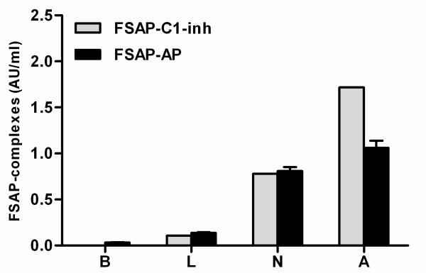
Figure 3.

FSAP complexes with C1-inhibitor and α2-antiplasmin as a measure of FSAP activation. Living (L), necrotic (N) and apoptotic (A) cells were incubated with r-plasma for 30 minutes at 37°C. Thereafter complexes of FSAP with either AP or C1-inh were measured by ELISA. R-plasma incubated with buffer without cells (B) was used as a negative control. Results were expressed in AU/ml. Plasma of 20 healthy donors was incubated with apoptotic cells for 30 minutes at 37°C. Complexes of FSAP with either AP or C1-inh were measured by ELISA. The median of the level of FSAP-inhibitor complexes in these controls was arbitrarily set as 0.5 AU/ml and used as a reference. Results are given as mean ± SEM, (n = 3).
