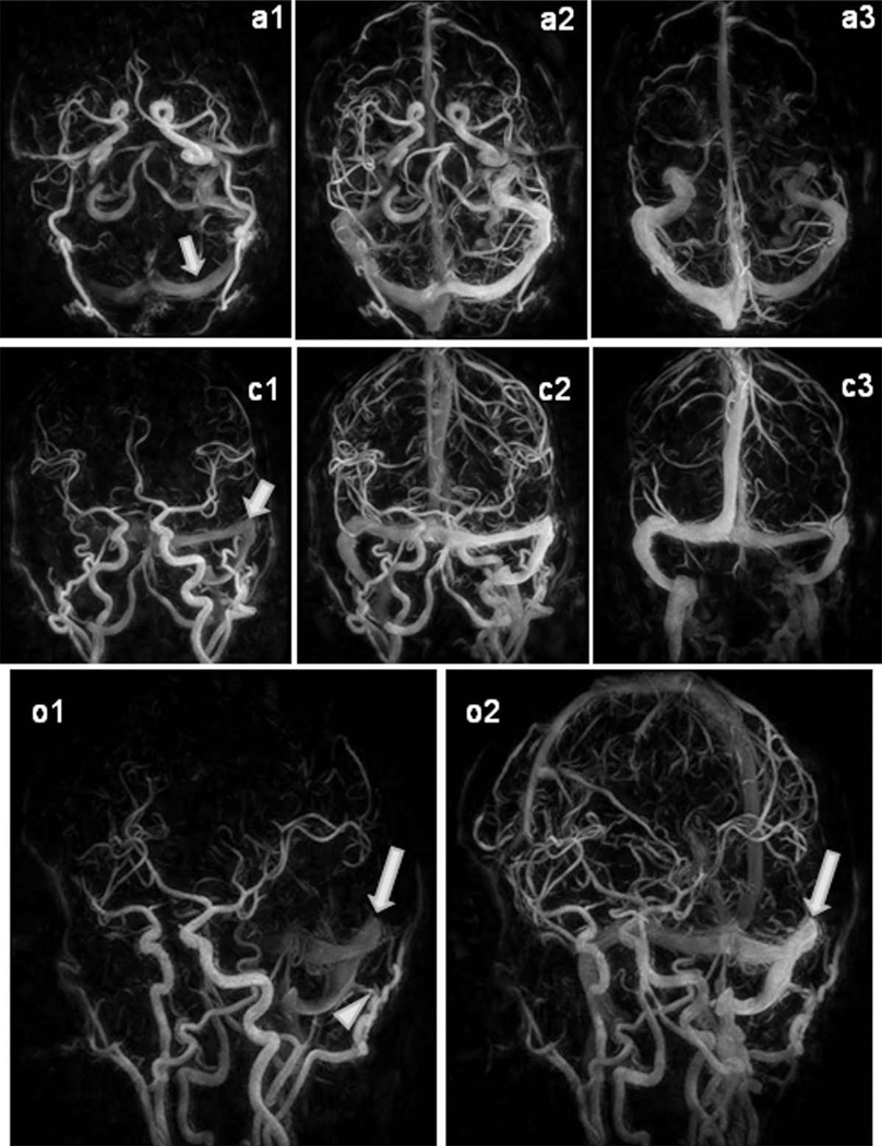
FIG. 4.
HYPR CE exam of a patient with dural Arteriovenous fistula. Arterial, mixed and venous phase images from the 60 frame HYPR CE exams are displayed in the axial, coronal and oblique views. Note the rapid enhancement of the left transverse sinus (arrows). On the oblique views a branch of the occipital artery supplying the dural arterio-venous fistula can be identified (arrowhead).

