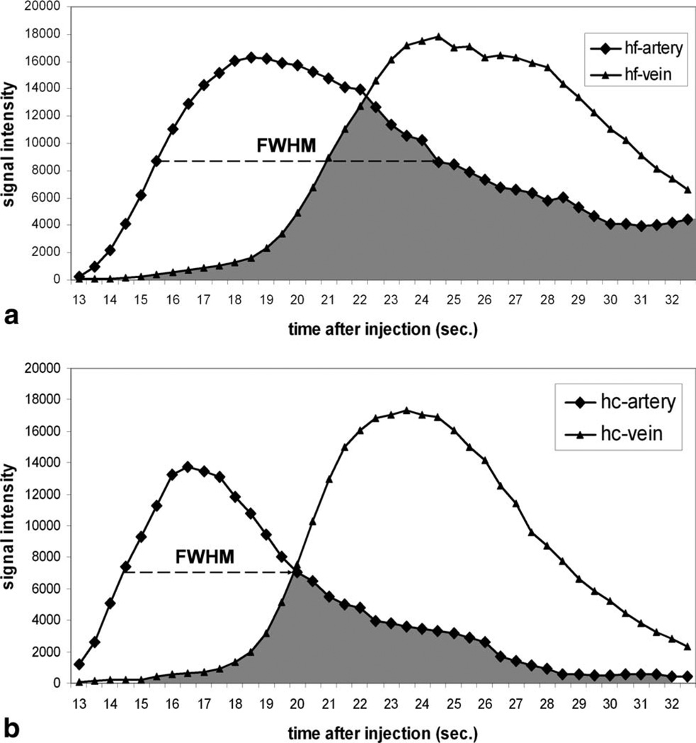
FIG. 6.
Example demonstrating the methods used to assess the arterial venous separation for (a) PC HYPRFlow and (b) HYPR CE. FWHM of the arterial curve from HYPR CE is significantly shorter than that from HYPRFlow. The overlap integral (shown as the shading area) from HYPR CE is significantly smaller than that from HYPRFlow.

