Fig. 3.
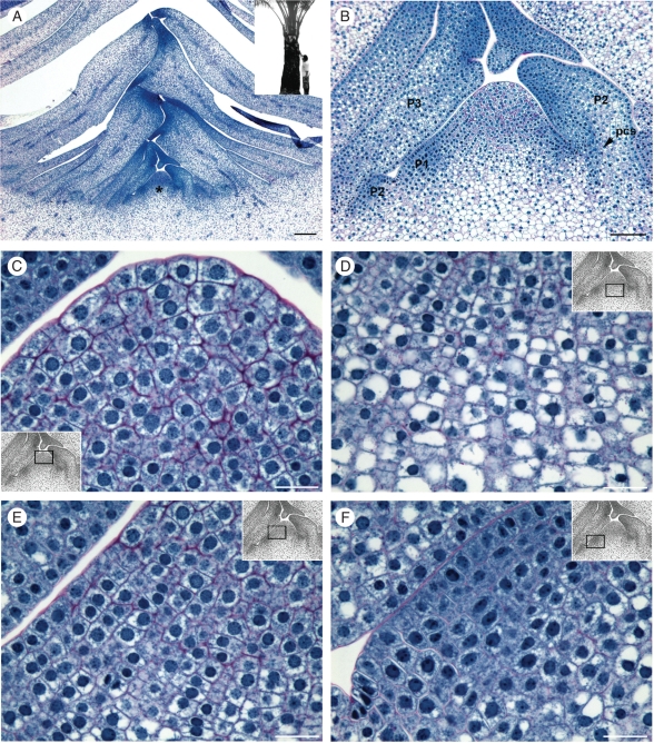
Shoot apex organization in a mature reproductive oil palm. (A) Shoot apex median longitudinal section from a 10-year-old plant. Inset: the corresponding plant used for shoot apex preparation. (B) Higher magnification view of (A) in the meristematic dome region. (C–F) Higher magnification views of the different histological zones of the meristematic dome shown in (B). (C) The central zone at the top of the meristematic dome – note the large cells with thick cell wall and central uncondensed nuclei. (D) The rib meristem zone and the parenchyma beneath the meristematic dome – note the progressive vacuolization and cell size enhancement. (E) The peripheral zone in the left part of the longitudinal section – note the anticlinal files of cells with a thinner cell wall and progressive vacuolization. (F) P1 leaf primordium at the early stage of protrusion – note the dense cytoplasm and nuclei, and limited vacuolization. The insets in (C–F) indicate the corresponding region of the SAM. Abbreviations: *, meristematic dome; P1–P2–P3, leaf primordia. Scale bars: (A) = 250 µm; (B) = 100 µm; (C–F) = 25 µm.

