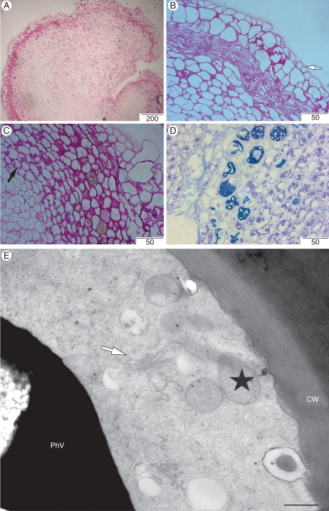
Fig. 8.
Histological and ultrastructural aspects of the callus sector. (A) General view of callus stained with PAS reagent. (B) Detailed view of A showing an epidermis-like cell layer (arrow). (C) Presence of amyloplast in the callus sector (black arrow). (D) Histological section of the callus sector in contact with the embryogenic sector revealing the accumulation of phenolic substances in the cells. (E) Detailed view of the phenol-storing cells showing numerous mitochondria (stars), and Golgi complex (arrow). Scale bars: (A) = 200 µm, (B, C) = 50 µm, (D) = 500 µm, (E) = 500 nm.

