
Figure 5.
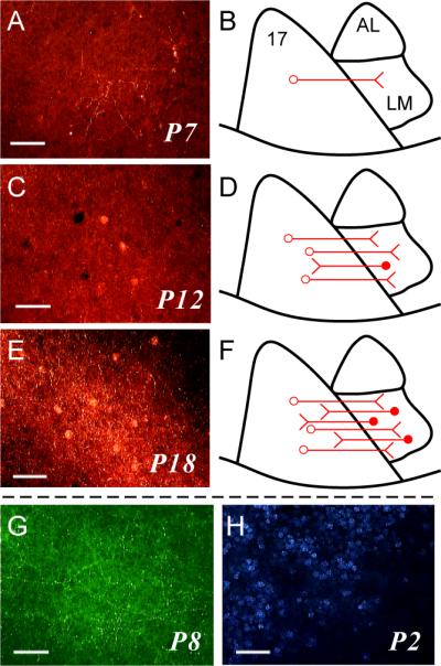
Development of feedforward and feedback connections between area 17 and area LM. A, C, E: Tangential sections through area LM after injections of DA-594 into area 17 at different postnatal days. B, D, F: Schematics summarizing the labeling pattern observed across cases at each time point. A,B: postnatal day 7 (P7). C,D: P12. E,F: P18. Anterograde label is present in all sections, indicating the presence of FF connections, but retrogradely labeled cell bodies (FB neurons) are present only at P12 and P18. G: Tangential section through area LM showing clear anterograde labeling and absence of retrograde labeling after an injection of BDA into area 17 at P8. H: Tangential section through region on border of LM showing callosally projecting neurons retrogradely labeled after an injection of bis-benzimide into the contralateral hemisphere at P2. Scale bars: 50 μm in all panels.