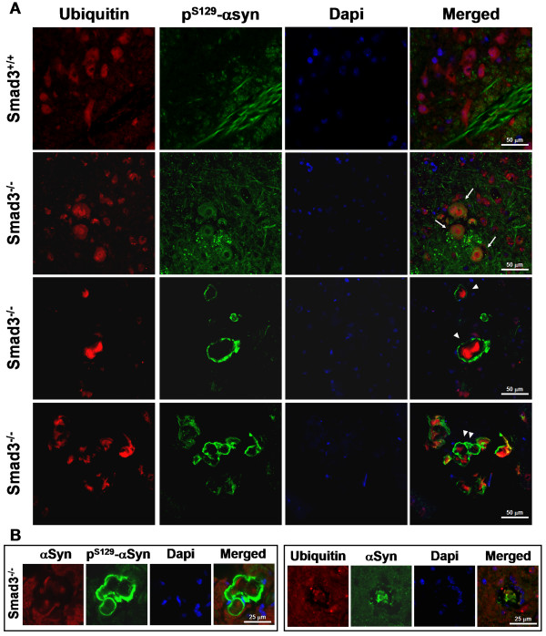
Figure 10.
α-synuclein deposits in Smad3 deficiency are ubiquitinated and S129 phosphorylated. (A) Confocal images of aggregates double-labelled with ubiquitin and PS129-α-synuclein in the ventral midbrain of Smad3 deficient mice. Different morphologies could be detected, from a clear core of ubiquitin surrounded by a halo of PS129-α-synuclein (arrows), to strong accumulation of ubiquitin and PS129-α-synuclein in isolated (arrowhead) or groups (double arrowhead) of deposits. (B) α-synuclein staining co-localize with ubiquitin within the core of the aggregates.

