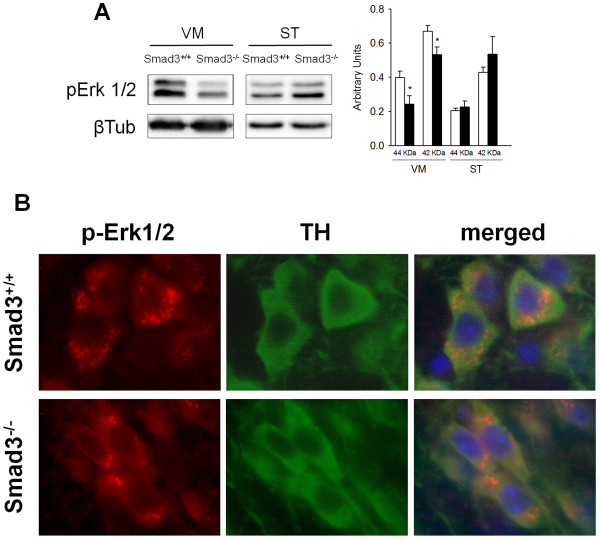
Figure 7.
Decreased Erk1/2 signalling in dopaminergic neurons of Smad3-/- mutants. (A) Analysis of the phosphorylation state of Erk1/2 MAPK in the VM and ST of Smad3 null mice identifies a specific decrease in Erk1/2 activation in the VM (*P < 0.05, Student's t-test, n = 6 per genotype, each measured two to three times). (B) Immunolabelling for p-Erk1/2 defines granular cytoplasmic structures in dopaminergic neurons that are fewer when Smad3 is depleted.

