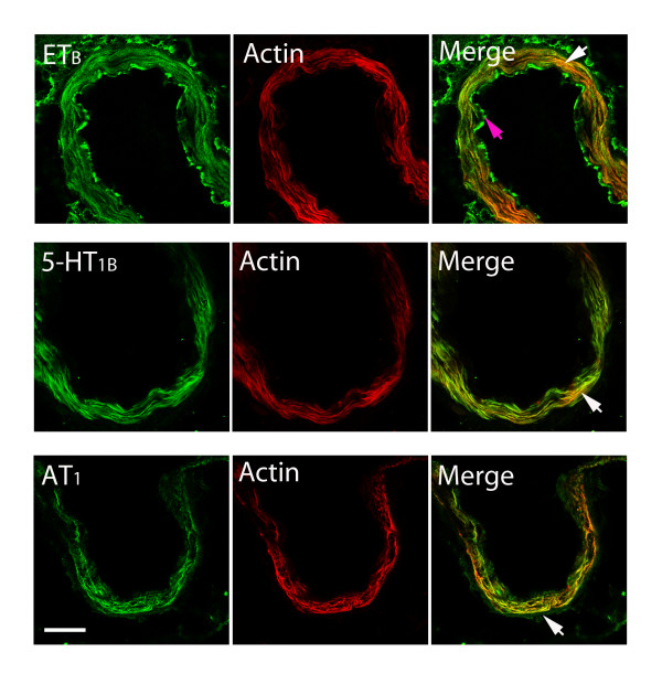
Figure 6.
Double immunofluorescence staining for ETB, 5-HT1B and AT1 and actin in smooth muscle cells of the basilar artery after SAH. Photographs demonstrating the localization of ETB, 5-HT1B, AT1, actin immunostaining, and their co-localization in smooth muscle cells (yellow fluorescence in the merged picture). Scale bar, 50 μm.

