
Figure 4.
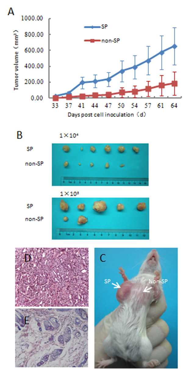
SP cells were more tumorigenic. (A) Tumor volumes (mean ± SEM) were plotted for 1 × 10³ cells of each population (SP, non-SP) injected (n = 6 per group). Tumors derived from SP were larger than those from non-SP. (B) Representative tumors due to injection of SP cells (1 × 10⁴ cells, 1 × 10³ cells) compared with non-SP injection (1 × 10⁴ cells, 1 × 10³ cells). (C) A representative tumor in a mouse specimen at the SP injection (1 × 10³ cells) site, but not at the non-SP injection (1 × 10³ cells) site. Histology from the SP injection site ((D), Original magnification, ×200) contained malignant cells, whereas the non-SP injection site ((E), Original magnification, ×200) revealed only normal mammary tissue.