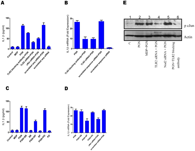
Figure 2. PGN mediated IL1β production is through TLR2 and involves NF-κB and JNK pathways.
(A-E), Blocking antibody (1 µg/ml) was added 20 minutes prior to stimulation, transfection with gene specific siRNA or scrambled sequence siRNA was done for 72 hours. MAPK inhibitors, SP600125 (20 µM) or PD98059 (50 µM) or SB202190 (5 µM) were added 20 minutes prior to stimulation (A) Normal macrophages or TLR2 knockdown macrophages or cRel knockdown macrophages or macrophages pre-treated with TLR2 specific blocking antibody were treated as indicated. After 12 hours culture supernatant was collected and the level of secreted IL1β was measured by ELISA. Each well contained 1×10⁶ macrophages. Statistical significance was checked by one way ANOVA. P value was found to be significant (<0.0001). (B) Real time RT-PCR analysis of IL1β mRNA. Normal macrophages or TLR2 knockdown macrophages or macrophages pre-treated with TLR2 specific blocking antibody were treated as indicated. Total RNA was isolated after 2 hours and used for checking IL1β transcripts. Each bar represents fold expression relative to untreated macrophages. Statistical significance was checked by one way ANOVA. P value was found to be significant (<0.0001). (C) Macrophages were left untreated or pre-treated MAPK inhibitors (JNK inhibitor SP600125 (20 µM) or ERK inhibitor PD98059 (50 µM) or p38 inhibitor SB202190 (5 µM)) for 30 minutes and then stimulated with MDP or PGN as indicated. Culture supernatants were collected after 12 hours and checked for the presence of secreted IL1β using ELISA. Each well contained 1×10⁶ macrophages. Statistical significance was checked by one way ANOVA. P value was found to be significant (<0.0001). (D) Real time RT-PCR analysis of IL1β mRNA. Macrophages were pre-treated with inhibitors or left untreated as described and stimulated with PGN or left unstimulated and checked for the presence of IL1β transcripts after 2 hours of stimulation. Each bar represents fold expression relative to untreated macrophages. Statistical significance was checked by one way ANOVA. P value was found to be significant (<0.0001). (E) Western blot analysis of phospho-cJun. TLR2 knockdown macrophages or Nod2 knockdown macrophages or macrophages pre-treated with TLR2 specific blocking antibody were stimulated as indicated. Cell extracts were collected after 40 minutes of stimulation and checked for the presence of phosphorylated form of cJun. Results in graph are presented as the mean of triplicate wells ± SD. Blots correspond to one representative experiment of three independent experiments.

