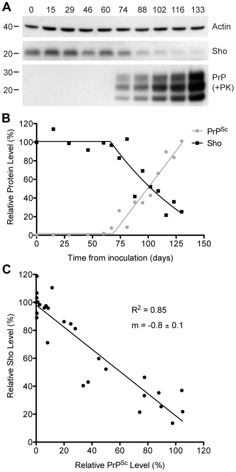
Figure 2. Inverse relationship between Sho and PrPSc levels during prion disease in mice.
(A) Western blotting of Sho and protease-resistant PrPSc in brain homogenates prepared from wt mice infected with RML prions at the indicated days postinoculation (dpi). As Sho signals began to decrease, protease-resistant PrPSc increased. Actin levels are shown as a control. Molecular masses based on the migration of protein standards are shown in kilodaltons. Sho and PrP were probed using the antibodies 06rSH-1 and HuM-P, respectively. (B) Quantification of relative Sho (black) and PK-resistant PrPSc (gray) levels in RML-infected mice at the indicated days postinoculation. The inflection points for Sho reduction and PrPSc accumulation both occurred at ∼70 dpi. (C) Correlation analysis of Sho and PK-resistant PrPSc levels in the brains of RML-infected mice (n = 29). A significant, inverse correlation (P<0.0001; R 2 = 0.85) was observed, indicating that increased protease-resistant PrPSc levels are associated with decreased Sho levels in the brain.

