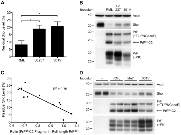
Figure 10. Decreased Sho levels correlate with the amount of PrPSc C2 fragment present in prion-infected animals.
(A) Quantification of Sho levels in meadow voles before and after infection with RML, Sc237, and 301V prions. Sho levels were reduced by ∼90% in RML-infected voles compared to the ∼80% reduction observed in Sc237- and 301V-infected voles (*P<0.05 as determined by one-way ANOVA, n = 3–4 for each group). (B) Western blot analysis of Sho levels in the brains of prion-infected meadow voles. Infection with RML prions resulted in the largest decrease in Sho levels and the highest amount of PrPSc C2 fragment (determined after digestion with thermolysin (TL) and PNGaseF). The presence of PK-resistant PrPSc indicates prion disease. (C) Correlation analysis of Sho and relative PrPSc C2 fragment levels in the brains of prion-infected meadow voles (n = 11). A significant, inverse correlation (P<0.001) was observed, indicating that increased production of the PrPSc C2 fragment is associated with decreased Sho levels in the brain. (D) Western blot analysis of Sho levels in the brains of Tg(MoSho)24474 mice infected with RML, Me7, and 301V prions. The largest decrease in Sho levels was observed with RML and Me7 infections, which also resulted in the largest amounts of PrPSc C2 fragment (determined after digestion with TL and PNGaseF). In comparison, infection with 301V prions resulted in the smallest reduction in Sho levels and the lowest relative level of the PrPSc C2 fragment. Sho and PrP were probed using the antibodies 06rSH-1 and HuM-P, respectively. Actin levels are shown for comparison. Molecular masses based on the migration of protein standards are shown in kilodaltons.

