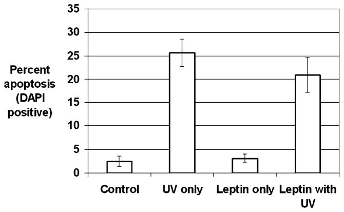
Fig. 4.
UV irradiation-induced stellate cell apoptosis detected by DAPI. Stellate cells were isolated from wild-type mice and plated on plastic coverslips for activation. After 7 days, stellate cells demonstrated morphological signs of activation. Stellate cells were incubated with control medium or 100 nM leptin for 24 hr. HSCs were exposed to UV irradiation at a dose of 30 J/m2. Six hours after UV irradiation, HSCs were fixed with 4% paraformaldehyde and stained with DAPI. Chromatin con was used to identify apoptotic stellate cells by DAPI, and apoptotic stellate cells were calculated as a percentage of total stellate cells. Results are shown as -fold increase in apoptosis compared to control baseline ± SE

