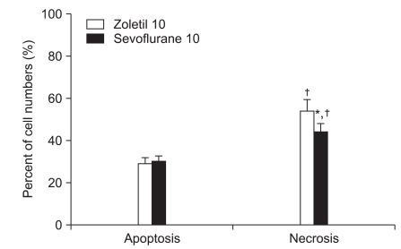
Fig. 2.
The percentage of necrotic and apoptotic cells in the hippocampus 7 days after forebrain ischemia. The percentage of necrotic cells was lower in the sevoflurane 10 min ischemia group (*P < 0.05). The percentage of necrotic cells in each anesthetic group is higher in the 10 min ischemia group compared with the 6 min ischemia group (†P < 0.05). There are no significant differences between groups with respect to apoptotic cell numbers. All data are expressed as mean ± SD.

