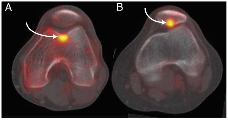
Figure 1.

A) Sample 18F NaF PET/CT image of the patellofemoral joint with a region of increased tracer uptake in the trochlea. B) Sample 18F NaF PET/CT image of the patellofemoral joint with a region of increased tracer uptake in the patella. Regions of increased tracer uptake are indicated by the arrows.
