
Fig. 4. RelA/p65 is responsible for DNMT-1 assembling on BRMS1 promoter.
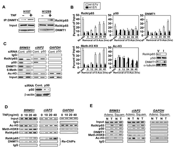
(A) TNF increased the interaction of RelA/p65 and DNMT-1. NSCLC cells were treated with TNF (40ng/ml) or not for 6 hrs. Co-IPs were performed using anti-DNMT-1 antibody, followed by immunoblotting for the presence of RelA/p65. (B) H157V/I cells were treated with 5-Aza as described above. ChIP analysis was performed at the indicated time points after removal of 5-Aza in H157 V/I cells by quantitative PCR specific to the BRMS1 promoter. Western blots show the expression of indicated proteins (nuclear RelA/p65, p50, DNMT1 and α-tubulin) in H157 V/I cells. (C) H157 cells were transiently transfected with siRNA control or p50. Post-transfection 48 hrs, ChIP analysis was performed across the BRMS1 promoter, and the promoters of cIAP2 and GAPDH. The siRNA transfection efficiency was proved by Western blot. (D) ChIP and re-ChIP analyses were performed in HEK 293T cells treated with TNF at the indicated doses for 16 hrs across the BRMS1 promoter, and the promoters of cIAP2 and GAPDH. Re-ChIP analysis used anti-RelA/p65 as the first immunoprecipitating antibody. (E) ChIP analyses were performed using patients’ samples (Adeno: adenocarcinoma, Squam: squamous cell cancer, T: lung tumor, N: matched adjacent non-cancerous lung tissue,) cross the BRMS1 promoter, as well as the promoters of cIAP2 and GAPDH.