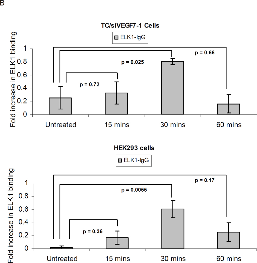
Figure 5. SDF-1α induces binding of the ELK-1 transcription factor to the pdgf-b promoter.
(A) Schematic of the pdgf-b promoter region with the potential ELK-1 binding sites identified using the GeneRegulation.com MATCH software. Two of the potential ELK-1 binding sites which scored >85% were investigated: at −600 bp (box 1) and at the TS site (box 2). Primer binding sites for the corresponding regions are also illustrated. (B) qPCR for the −600 bp site; the fold increase in ELK-1 binding is scaled from 0 (non-binding) to 1 (complete binding). TC/siVEGF7-1 and HEK293 cells were plated, cultured in the absence of growth factors and supplements for 24 hours, and then treated with 100 ng/mL of SDF-1α for 15, 30, or 60 minutes. The cells were collected and ELK-1 binding quantified by ChIP. Each bar graph shows pooled data from 3 independent experiments, each performed in triplicate, +/− SD. The sample values minus the control values were used in one-way analysis of variance. Negative values were replaced with 0, indicating non-binding. P < 0.05 was considered significant.


