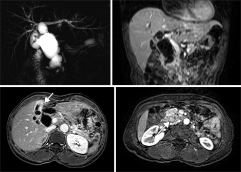
Fig. 1.
Magnetic resonance cholangiopancreatography (MRCP) image including vertical and horizontal sections. MRCP shows fusiform cystic changes involving the entire common bile duct and hilar bile duct compatible with a choledochal cyst (type Ia) associated with the long common channel of the pancreaticobiliary junction (C-P type). Irregular wall thickening at the fundus and body of the gallbladder was seen. A 2.5 × 1.9 cm mass is also seen around the pancreaticobiliary junction.

