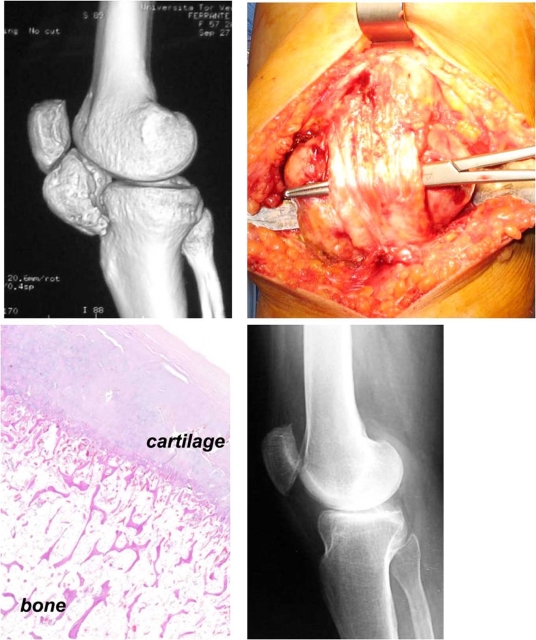
Fig. (1).
Three-dimensional CT scan examination of the right knee in a 58-year-old woman affected by a giant osteochondroma located between the apex of the patella and the proximal tibia (A). The tumor was isolated behind the patellar tendon and marginally resected (B). At histological examination, the bone mass formed by endochondral ossification was surrounded by an outer layer of hyaline cartilage without chondrocyte atypia (C). At follow-up, 8 years after surgery, the radiographic examination did not show any evidence of recurrence (D).

