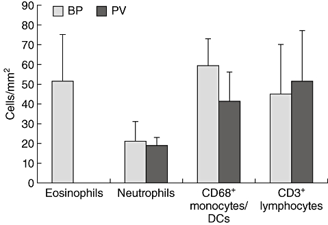
Fig. 3.

Eosinophils predominate bullous pemphigoid (BP) lesions. Cells were stained by immunofluorescence and counted per visual field. Numbers were calculated per mm2. The average densities and standard deviations of five different subepidermal tissue areas of nine individual patients with BP and four patients with pemphigus vulgaris (PV) for each cell type are shown.
