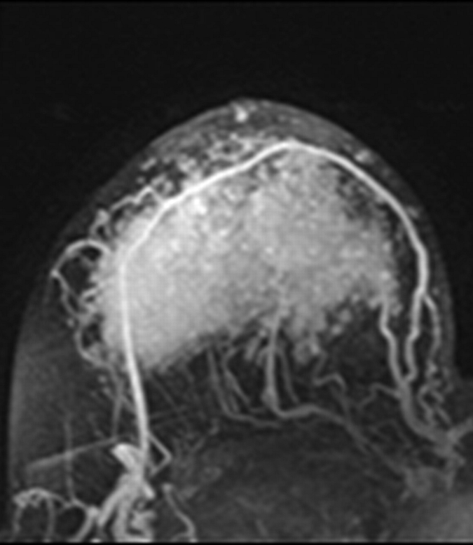
Figure 2a:

Axial (a–c) maximal intensity projection and (d–f) original subtraction MR images in 32-year-old woman with HER2-negative hormone receptor–positive Ki-67 20% breast cancer in right breast. On (a, d) baseline images obtained before NAC, the tumor appears as a 9.8-cm lesion with diffuse, nonmasslike enhancement. On (b, e) images obtained during NAC, the tumor is reduced to 5.6 cm, and on (c, f) images obtained after NAC, the tumor is reduced to 3 cm. Pathologic examination showed scattered cancer nests in a 10-cm area.
