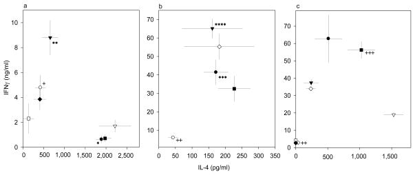
Figure 5.
Modification of OVA-tg T-cell differentiation with spleen cells from stat4−/− DO11.10+/− (a), stat6−/− DO11.10+/− (b), or wild type DO11.10+/− (c) mice. Cells were differentiated with OVA in the absence of various additives (black circle) or presence of TGF-β (open circle), anti-IL-4 (black triangle), anti-IL-12 (open triangle), Pb (black square), TGF-β+anti-IL-4 (open square), TGF-β+anti-IL-12 (black diamond), or Pb+anti-IL-4 (open diamond). Data were obtained from 6 mice assayed individually in six separate experiments, and the results are expressed as the mean amounts (± SEM) of IL-4 and IFN-γ produced.

