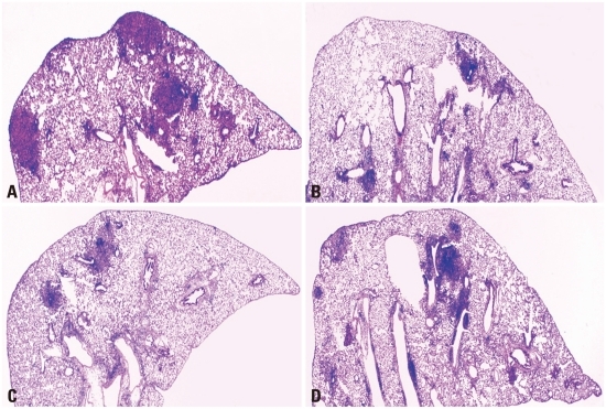
Fig. 4.
Histopathology of lungs after aerosol infection with virulent M. tuberculosis. The right middle lobe of the lungs of mice was removed after 90 days post-challenge and lung sections were stained with H&E (×20). The representative histopathology of lungs of naïve mice (A), the BCG vaccine-immunized mice (B), co-immunized mice of the BCG vaccine and IL-12 DNA vaccine (C), and IL-18 DNA vaccine (D).

