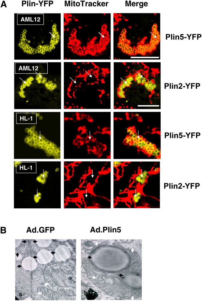
Fig. 5.
Plin5 recruits mitochondria in a cell type-independent manner. (A) Murine AML12 cells (liver; top row) and HL1 cells (heart; bottom row) were transfected with Plin5-YFP or Plin2-YFP and incubated with 400 μM oleic acid overnight. The following day, cells were incubated with MitoTracker (1 μM) for 30 min, and confocal microscopy was performed as in Fig. 1. Micrographs show merged image obtained for Plin-YFP and MitoTracker in representative cells from two to three separate experiments. Bar: 10 μm. White arrows indicate LDs. (B) AML12 (liver) cells were transduced with GFP-adenovirus or Plin 5-YFP adenovirus and TEM was performed. White arrows indicate mitochondria, black arrows indicate LD. Magnification: 10,000×.

