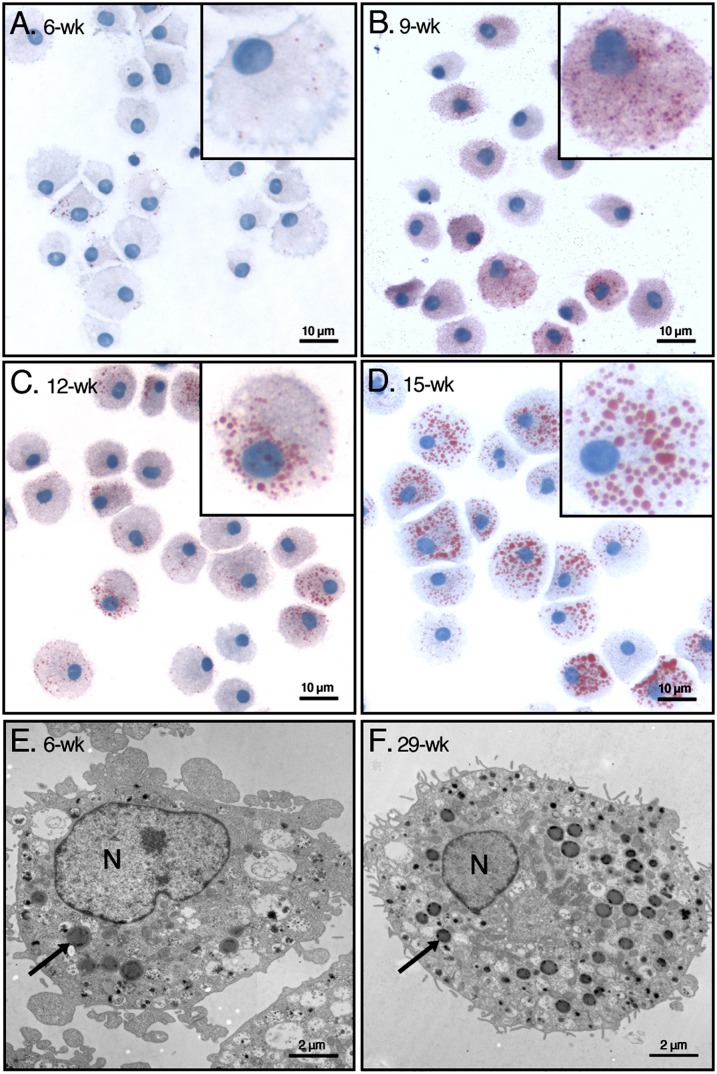
Fig. 1.
Identification of LBs in human MCs with light and with electron microscopy. Human peripheral blood-derived CD34+ progenitor cells from 18 donors were grown under defined culture conditions, which induced their differentiation into mature MCs, as described in Materials and Methods. A–D: The cells were stained with Oil Red O/hematoxylin at the indicated weeks in culture. Representative images of cells from a single donor are shown. Each inset shows one typical cell representative for the corresponding time point in culture. E, F: Mast cells from an early (6-week) and late (29-week) culture were analyzed by transmission electron microscopy. In each panel, one typical LB is indicated with an arrow. N, nucleus; wk, week.

