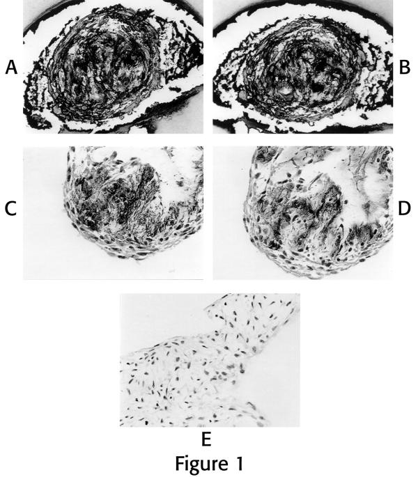
Figure 1.
Photomicrographs of ECM immunolocalizations of cells grown in the 3D microenvironment: Figures 1A and B are serial sections through a multi-celled colony evaluated for localization of chondroitin sulfate (Fig. 1A), keratan sulfate (Fig 1B). Figures 1C and D are serial sections through a multi-celled colony evaluated for localization of Type I collagen (Fig. 1C), and Type II collagen (Fig. 1D). Note positive black localization product for these ECM components. Figure 1E is a photomicrograph from a different specimen which was used as the negative control with deletion of primary antibody for this series of localizations with hematoxylin staining. (All photomicrographs X 295). Cells were from the same subject, were first passage and were grown for 10 days.

