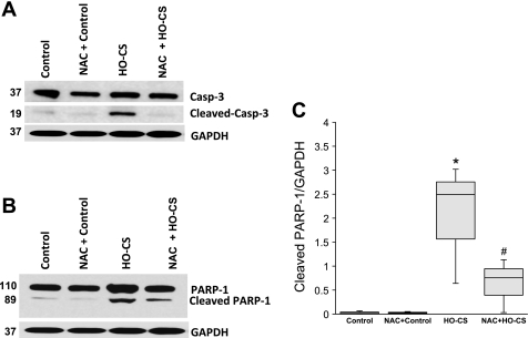
Fig. 5.
Cleaved caspase-3 (A) and its downstream substrate PARP-1 (B) in mouse lung epithelial cells (MLE-12) subjected to control (lane 1), NAC + control (lane 2), HO-CS (lane 3), and NAC + HO-CS (lane 4). Significant cleavage of caspase-3 and PARP-1 was observed in HO-CS. In NAC + HO-CS, activation of caspase-3 was completely inhibited. C: box plots of cleaved PARP-1 densitometry show NAC + HO-CS significantly reduced cleaved PARP-1 compared with HO-CS. Horizontal lines indicate median, shaded boxes indicate inner quartile, and whiskers indicate data range. Representative blots and densitometry were from 3 independent experiments in which 4 wells were pooled for each condition. *P < 0.05 compared with control. #P < 0.05 compared with HO-CS.

