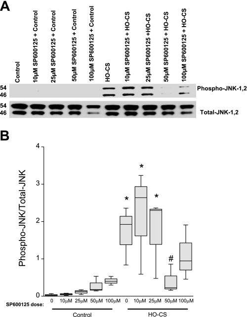
Fig. 7.
Pharmacological inhibition of JNK phosphorylation by SP600125 (A) in MLE 12 cells. n = 3 Independent experiments in which 4 wells were pooled for each condition. A: SP600125 treatment in control cells (lane 1), control cells in the presence of several doses of SP600125–10 μM (lane 2), SP600125–25 μM (lane 3), SP600125–50 μM (lane 4), and SP600125–100 μM (lane 5); HO-CS without SP600125 (lane 6), and HO-CS with several doses of SP600125 pretreatment SP600125–10 μM (lane 7), SP600125–25 μM (lane 8), SP600125–50 μM (lane 9), and SP600125–100 μM (lane 10). B: box plot of densitometry of the Western blot in A shows that the 50 μM dose of SP600125 significantly inhibited JNK phosphorylation. Horizontal lines indicate median, shaded boxes indicate inner quartile, and whiskers indicate data range. *P < 0.05 compared with control. #P < 0.05 compared with HO-CS.

