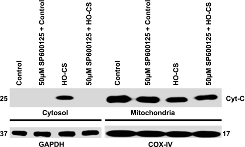
Fig. 8.
Cytochrome c release from mitochondria in MLE-12 cells subjected to control, SP600125 + control, HO-CS, and SP600125 + LIC. Cytoplasmic and mitochondrial fractions were separated, and Western blotting performed. Top: cytoplasmic fractions of control (lane 1), SP600125 + control (lane 2), HO-CS (lane 3), and SP600125 + HO-CS (lane 4); mitochondrial fractions of control (lane 5), SP600125 + control (lane 6), HO-CS (lane 7), and SP600125 + HO-CS (lane 8). Bottom: GAPDH was the loading control for cytoplasmic fractions (lanes 1–4), and COX IV was the loading control for mitochondrial fractions (lanes 5–8). n = 3 Independent experiments, and 4 wells were pooled for each condition. Cytochrome c release was observed in HO-CS, whereas SP600125 + HO-CS completely blocked cytochrome c release from mitochondria. SP600125 dose was 50 μM final concentration based on dose-response data.

