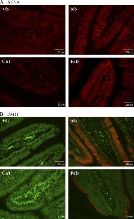
Fig. 5.
Immunohistochemical analysis of Atp7a and Dmt1 protein expression in rat duodenum. Fixed tissue sections were reacted with the anti-Atp7a- or -Dmt1-specific antiserum followed by a fluorescent-tagged secondary antibody and imaged with a confocal microscope. A: Atp7a protein is depicted by the red color. B: autofluorescence is shown (green) along with the specific signal (red color) depicting the Dmt1 protein. The confocal settings remained constant across all images. Images are typical of several experiments. Ctrl, control Sprague Dawley (SD) rat; FeD, iron-deficient SD rat.

