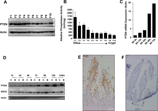
Fig. 2.
PTEN expression is induced during intestinal cell differentiation. A: PTEN protein expression in cells sequentially isolated along the mouse crypt-villus axis, with F1 corresponding to cells isolated from the villus tip and F10 corresponding to cells from the crypt base. B: confirmation of the fractionation efficiency as assessed by measurement of activity of the intestinal cell differentiation marker alkaline phosphatase in fractions 1–10. C and D: induction of PTEN mRNA (C) and protein (D) expression in Caco-2 cells treated with 5 mM butyrate. Shown in parallel is induction of the known differentiation marker keratin 8. E: immunohistochemical localization of PTEN in human small intestine. F: negative control (no primary antibody).

