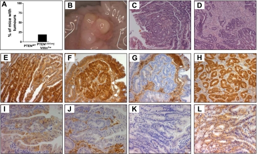
Fig. 5.
A: frequency of adenoma formation in PTENWT and PTENLox/Lox/villinCre mice at 12 mo of age. B: macroscopic image of a cecal tumor in a PTENLox/Lox/villinCre mouse. C and D: H&E staining demonstrating a tubulovillous adenoma (C) and an adenocarcinoma (D) derived from PTENLox/Lox/villinCre mice. E and F: tumors derived from PTENLox/Lox/villinCre mice show evidence of nuclear β-catenin. Tumors from PTENLox/Lox/villinCre mice were stained with β-catenin as indicated in methods. Nuclear β-catenin is indicated by arrows in F. G: negative control. Nonspecific staining is due to the use of an anti-mouse secondary antibody. Shown for comparison in H is nuclear β-catenin in an adenoma derived from an Apc1638 mouse that harbors an inactivating mutation at codon 1638 of the Apc gene. I and K: pAKT staining in adenomas derived from PTENLox/Lox/villinCre and Apc1638 mice, respectively. J and L: PTEN staining in adenomas derived from PTENLox/Lox/villinCre and Apc1638 mice, respectively. J: PTEN staining in stromal tissue shows specificity of PTEN deletion to epithelial cells.

