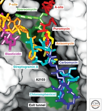
Figure 6.
The positions of seven antibiotics and A-site (red) plus P-site (yellow) substrates bound to the peptidyl transferase center. The ribosome has been split open to reveal the lumen of the exit tunnel and adjacent regions of the peptidyl transferase site. Ribosomal components are depicted as a continuous surface that is colored green at two positions where splayed out bases provide hydrophobic binding sites for small molecules. Seven independently determined cocrystal structures have been aligned by superimposing the 23S rRNA in each complex.

