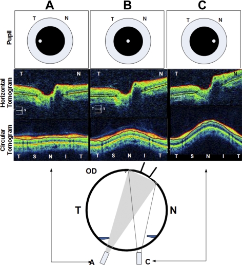
Figure 2.
The parallax artifact of the OCT in the right eye of a normal subject. Top row: location of the scanning beam on the pupil. The corresponding horizontal (second row) and circular (third row) tomograms are shown. When the aiming beam is located over the nasal region of the pupil in an emmetropic patient, the image is tilted (as in column C). To obtain a symmetrical untilted view of the disc and peripapillary region the beam must scan perpendicular to the target (column A). When the scanning beam is obliquely oriented (C, bottom inset), the echo delay of the nasal retina is shorter than the temporal retina, causing the image to tilt. The degree of tilting relative to beam location on the pupil may vary with the axial length, refractive error, and shape of the eye. T, temporal; N, nasal.

