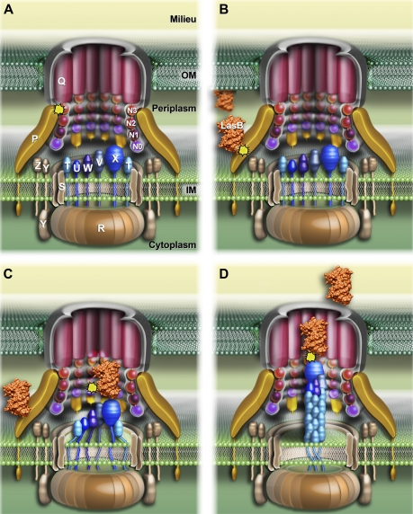
FIGURE 1.
Schematic three-dimensional model of the P. aeruginosa Xcp secreton showing substrate recognition and transport during the type II secretion process. A, the schematic representation of the P. aeruginosa Xcp secreton. All Xcp components (labeled following Xcp nomenclature only) are represented according to their cellular localization, topology, and multimerization state. B–D, the different consecutive steps followed by the substrate for its recruitment, transport, and release by the secreton during the type II secretion process (see “Discussion” for details). The interactions identified in this study are represented by yellow asterisks.

