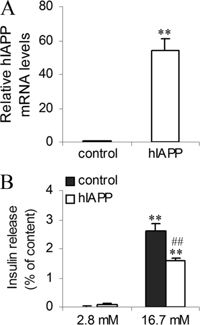
FIGURE 3.
Characterization and secretory capacity of islets overexpressing hIAPP. Islets were cultured in RPMI medium at 5.5 mm glucose. A, hIAPP mRNA levels of islets overexpressing hIAPP (hIAPP islets) were detected by real-time PCR. The data were normalized to GAPDH mRNA and presented as relative to control islets infected by pLenti-LacZ (control islets). **, p < 0.01 versus control islets (n = 3). B, 2 h before analyses, islets were depleted without glucose, after which insulin release was measured following 1.5 h of incubation at basal 2.8 mm glucose and stimulatory 16.7 mm glucose. Insulin secretion was tested in islets infected by pLenti-LacZ or pLenti-hIAPP. Values were normalized to total cellular content of insulin. **, p < 0.01 versus secretion at 2.8 mm glucose; ##, p < 0.01 versus corresponding control (n = 3). Results are expressed as mean ± S.E.

