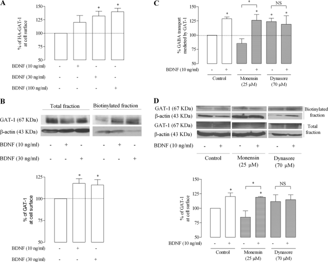
FIGURE 5.
BDNF enhances surface expression of GAT-1 in astrocytes. A, HA-rGAT-1-transduced astrocytes were incubated for 10 min with (+) or without (−) BDNF, as indicated below each bar and then assayed by ELISA. 100% on the ordinate represents normalized HA-GAT-1 expression in plasma membrane of astrocytes in the control situation (absence of BDNF). B, rGAT-1-transduced astrocytes were incubated as in A, but changes in surface GAT-1 immunoreactivity were assessed by surface biotinylation. In the upper panel is shown a representative immunoblot from total lysate and biotinylated astrocyte fractions. Blots were probed with anti-GAT-1 (1:500), and β-actin (1:10000) immunoreactivity was used as loading control. In the lower panel is shown the average densitometric analysis, where 100% on the ordinate represents normalized rGAT-1 expression in the biotinylated fraction in the absence of BDNF. C and D, influence of monensin and dynasore (for details, see “Results”) upon the effect of BDNF on GABA uptake (C) or surface expression of rGAT-1 (D). In the upper panel in D is shown a representative immunoblot from total lysate and biotinylated astrocyte fractions, and in the lower panel is shown the average densitometric analysis, where 100% on the ordinates represents normalized rGAT-1 expression in the biotinylated fraction in the absence of BDNF. The results are expressed as mean ± S.E. (error bars) from four (A), five (B and C), or three (D) individual experiments. *, p < 0.05 (one-way ANOVA followed by the Bonferroni's post-test), as compared with control conditions (first column on the left) except where otherwise indicated by the connecting lines above the bars. NS, not statistically significant (p > 0.05).

