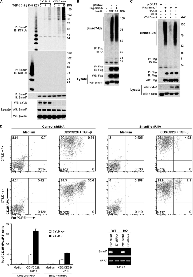
FIGURE 5.
Smad7 ubiquitination in response to TGF-β is enhanced in CYLD-deficient cells. A, Smad7 polyubiquitination in TGF-β-stimulated splenic wild type or CYLD-deficient CD4+ T cells was analyzed by immunoprecipitation (IP) of the proteins from denatured cellular lysates followed by immunoblotting (IB) with an anti-Lys-63-linked ubiquitin Ab (K63 Ub) or anti-K48-linked ubiquitin Ab (K48 Ub). The amount of immunoprecipitated Smad7 was determined by immunoblotting with anti-Smad7. WB, Western blotting; MW, molecular weight markers. B, cell lysates from HEK 293 cells transfected with pcDNA3, FLAG-Smad7, HA-ubiquitin, or HA-tagged Lys-63-only ubiquitin as indicated were immunoprecipitated as in A. The amount of polyubiquitinated FLAG-Smad7 in the immunoprecipitate was determined by immunoblotting with anti-HA, and the amount of immunoprecipitated FLAG-Smad7 was determined by immunoblotting with anti-FLAG. FLAG-Smad7 and β-actin levels in the lysate were determined by Western blotting. C, HEK 293 cells were transfected with pcDNA3, FLAG-Smad7, HA-tagged wild type ubiquitin, CYLD, and CYLD-mut lacking enzymatic activity as indicated. The cells were harvested, and the lysates were immunoprecipitated as in A. The amount of polyubiquitinated FLAG-Smad7 in the immunoprecipitate was determined by immunoblotting with anti-HA, and the amount of immunoprecipitated FLAG-Smad7 was determined by immunoblotting with anti-FLAG. The amounts of FLAG-Smad7, CYLD, and β-actin in the lysate were determined by Western blotting. D, transduced wild type and CYLD-deficient CD4+CD25− T cells were cultured with medium alone or in the presence of plate-bound anti-CD3/CD28 antibodies with TGF-β. At the end of the culture period, the cells were analyzed for CD25 and Foxp3 expression by flow cytometry. The experiment was repeated three times. Bottom left panel, bar graph demonstrating the mean and distribution of CD25+ Foxp3+ T cells. Bottom right panel, RT-PCR analysis of Smad7 expression in both WT and Cyld−/− primary CD4+CD25− T cells transduced with non-targeted or Smad7 (S7)-targeted shRNA.

