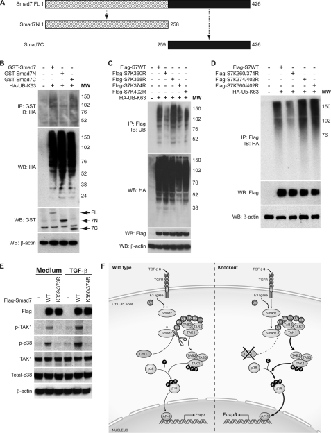
FIGURE 6.
Lys-63-linked polyubiquitination of Smad7 at its C terminus regulates TAK1 and p38 MAPK activities in response to TGF-β. A, schematic representation of the structure of wild type Smad7 and N- and C-terminal truncation mutants. B, cell lysates were prepared from HEK 293 cells transfected with GST-tagged Smad7, GST-tagged Smad7 deletion mutants (GST-Smad7N and GST-Smad7C), and HA-tagged Lys-63-only ubiquitin (HA-UB-K63) as indicated. The lysates were subjected to immunoprecipitation (IP) as in Fig. 5A. The amount of polyubiquitinated GST-Smad7 in the immunoprecipitate was determined by immunoblotting (IB) with anti-HA antibody. The amounts of ubiquitin, GST-Smad7, and β-actin in the lysate were determined by Western blotting (WB). MW, molecular weight markers. C, cell lysates from HEK 293 cells transfected with FLAG-Smad7, FLAG-Smad7-K360R (Flag-S7K360R), FLAG-Smad7-K368R (Flag-S7K368R), FLAG-Smad7-K374R (Flag-S7K374R), FLAG-Smad7-K402R (Flag-S7K402R), and HA-tagged Lys-63-only ubiquitin (HA-UB-K63) were subjected to immunoprecipitation as in Fig. 5A. The amount of polyubiquitinated FLAG-Smad7 in the immunoprecipitate was determined by immunoblotting with anti-HA. The amounts of ubiquitin, FLAG-Smad7, and β-actin in the lysate were determined by Western blotting. D, cell lysates from HEK 293 cells transfected with FLAG-Smad7, FLAG-Smad7-K360R/K374R (Flag-S7K360/374R), FLAG-Smad7-K374R/K402R (Flag-S7K374/402R), FLAG-Smad7-K360R/K402R (Flag-S7K360/402R), and HA-tagged Lys-63-only ubiquitin (HA-Ub-K63) as indicated were harvested and immunoprecipitated as in Fig. 5A. The amount of polyubiquitinated FLAG-Smad7 in the immunoprecipitate was determined by immunoblotting with anti-HA. FLAG-Smad7 and β-actin in the lysate were determined by Western blotting. E, HeLa Smad7 knockdown cells were reconstituted with FLAG-Smad7 or FLAG-Smad7-K360R/K374R (K360/374R). Following TGF-β stimulation, phospho-TAK1 (p-TAK1) and phospho-p38 (p-p38) levels were determined by immunoblotting. K359/373R, K359R/K373R. F, proposed model of the role of CYLD in TGF-β signaling and Treg development. TGFR, TGF receptor; Ub, ubiquitin; P, phosphorylated.

