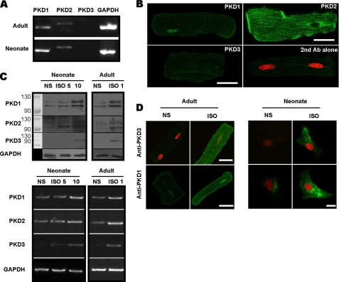
FIGURE 1.
The presence of PKD isoforms and their induction by ISO in cardiac myocytes. A, RT-PCR detection of mRNAs for PKD1 and PKD2 but not PKD3 in isolated adult (upper) and cultured neonatal (lower) myocytes. GAPDH was used as internal control. B, immunolocalization of each PKD isoform in adult myocytes as detected using a confocal microscopy. Cells were fixed and incubated with primary antibodies followed by reaction with Alexa-488 conjugated secondary antibody (green). Propidium iodide (PI) was used as a counterstain for nuclei (red). Note the fluorescent signal conferred by anti-PKD3 is as weak as that stained with second antibody alone, indicating PKD3 is absent in the adult myocyte. Scale bars = 25 μm. C, Western blot (upper) and RT-PCR (lower) assays showing expression up-regulation of three PKD isoforms by ISO in adult and neonatal myocytes. Adult cells were treated with 1 μm ISO for 24 h and neonate was treated with 5 and 10 μm ISO for 48 h. GAPDH was used as loading control. D, expression up-regulation of PKD3 and PKD1 by ISO in adult and neonatal myocytes as detected with a confocal microscopy. Immunofluoresent procedures were the same as that described in B. Scale bars, 25 μm.

