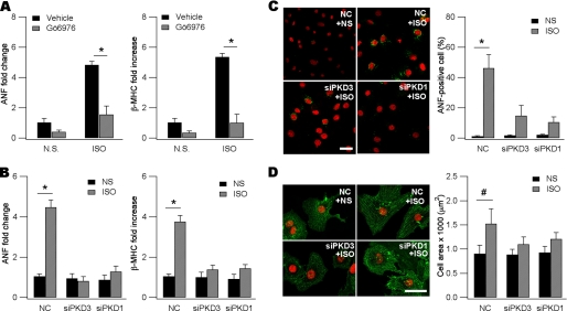
FIGURE 2.
PKD3 and PKD1 are both required for ISO-induced fetal gene reactivation and hypertrophy. A, induction of ANF (left) and βMHC (right) mRNAs by ISO (10 μm, 48 h) was blunted by 2 μm Gö 6976 (n = 3, *, p < 0.01, ANOVA). Gö 6976 was dissolved in DMSO (vehicle) as stock in 2 mm, applied 30 min before ISO treatment, and remained in the culture throughout the experiment. mRNAs were detected with qPCR and 18 S rRNA as an internal standard. The mean value for expression of each gene in DMSO-treated cells is defined as 1. NS, normal saline. B, ISO-induced ANF (left) and βMHC (right) mRNAs were prevented by gene silencing of PKD3 and PKD1 (n = 3, *, p < 0.01, ANOVA). Note in the absence of ISO, siPKD3, or siPKD1 did not affect baseline ANF and βMHC. Cells were transfected with negative control (NC) siRNA, siPKD3, or siPKD1 followed by NS or ISO application. The mean value for mRNAs of each gene in NS- and NC-treated cells is defined as 1. C, ISO-induced ANF expression was reduced by knockdown of PKD3 or PKD1. Cells were fixed and incubated with anti-ANF followed by reaction with Alexa-488 conjugated second antibody (green). Shown on the right panel is quantitative analysis (*, p < 0.01, Pearson's chi-square test). Data are expressed as means ± S.D. For each experiment at least 50 cells were counted. Scale bar, 25 μm. D, ISO-induced cardiac myocyte hypertrophy was blunted by gene silencing of PKD3 or PKD1. Cells were fixed and incubated with anti-α-actinin followed by reaction with Alexa-488-conjugated second antibody (green). Note larger cell size and highly- organized sarcomeres in ISO-treated myocytes. In the absence of ISO, siPKD3, or siPKD1 did not affect baseline cell morphology. Scale bar, 25 μm. Shown on the right panel is quantitative analysis (n = 75–130, #, p < 0.05, ANOVA). Data are expressed as means ± S.D.

