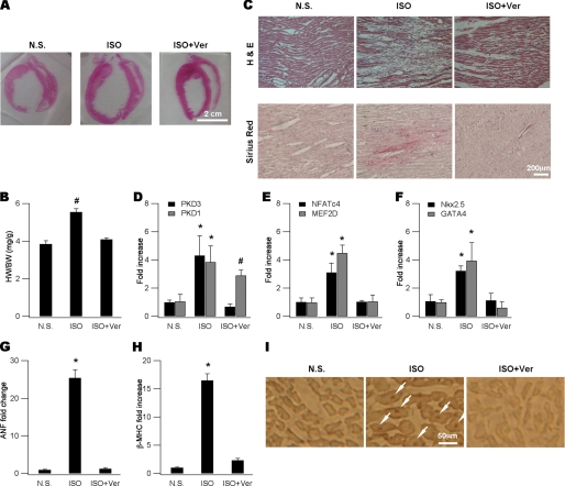
FIGURE 4.
PKD3-mediated hypertrophic signaling in in vivo cardiac hypertrophy. A, representative gross heart pictures showing ISO-induced hypertrophy and its blockade by verapamil (Ver). ISO was injected at a dose of 5 mg kg−1 d−1 and Ver (10 mg kg−1 d−1) was administrated by dissolving in drinking water. Animals were sacrificed on day 7 and heart sections were made for histology and immunohistochemistry. Isolated ventricular myocytes were made for mRNA determination. B, comparison of HW/BW ratios (mg/g) for animals shown in (A) (n = 5, #, p < 0.05 versus N.S., ANOVA). C, ISO injection resulted in prominent cell loss (upper, H and E) replaced with fibrosis (lower, Sirus Red) and these were attenuated by verapamil. D, mRNAs of PKD3 and PKD1 were increased by isoproterenol and prevented by co-administration with verapamil (n = 5, *, p < 0.01, #, p < 0.05 versus N.S., ANOVA). E and F, mRNAs of NFATc4, MEF2D (E), GATA4 and Nkx2.5 (F) were elevated by isoproterenol and prevented by co-administration with verapamil (n = 5, *, p < 0.01 versus N.S., ANOVA). G and H, mRNAs of ANF (G) and βMHC (H) were up-regulated by isoproterenol and prevented by co-administration of verapamil (n = 5, *, p < 0.01 versus N.S., ANOVA). I, in situ detection of nuclear NFATc4 proteins (arrows in the middle panel) using immunohistochemistry in an isoproterenol-treated animal and this was not observed in verapamil-coapplied animals (right).

