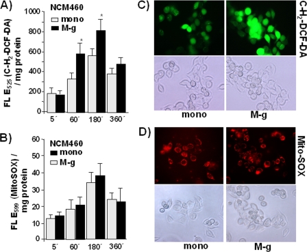
FIGURE 3.
The exposure to inflammatory macrophages increases ROS level in NCM460 cells. After labeling with 10 μm cH2DCF-DA (A) or 250 nm MitoSOXTM (B) for 30 min at 37 °C, NCM460 cells were either mono or cocultured (M-g) for the indicated periods. Then fluorescence was determined; the data represent the means ± S.D. of four independent experiments. *, p < 0.05 compared with the corresponding monocultured cells. In parallel, living cell fluorescence microscopy of NCM460 cells stained with 1 μm cH2DCF-DA (C) or 250 nm MitoSOXTM (D) for 10 min at 37 °C followed by mono or coculture for 1 h. Images of 40× magnification and representative results of two independent experiments are shown.

