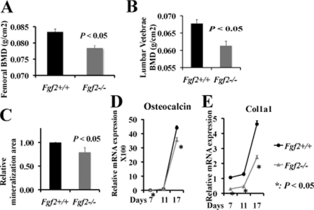
FIGURE 1.
Reduced bone formation and decreased osteoblast differentiation in Fgf2−/− female mice compared with wild-type littermates. Femoral BMD (A) and lumber vertebrae BMD (B) were decreased significantly in Fgf2−/− mice. Reduced osteoblast mineralization (C), reduced osteocalcin (D), and Col1a1 (E) mRNA expression in Fgf2−/− compared with Fgf2+/+ BMSCs are shown. (Black line represents Fgf2+/+, and gray line represents Fgf2−/−). A and B, BMD was determined in 6-month-old female Fgf2+/+ (n = 7) and Fgf2−/− (n = 8) mice. C, freshly isolated BMSCs were cultured in α-MEM/10% FBS for 9 days, then in α-MEM/10% FBS with 50 μg/μl ascorbic acid and 8 mm β-glycerophosphate for another 8 days, followed by alkaline phosphatase, von Kossa staining, and quantification of total mineralization area. Data are mean ± S.E. of four independent experiments using 8-month-old male mice. D and E, freshly isolated BMSCs from 12-month-old female mice were cultured in α-MEM/10% FBS for 3 days, then in α-MEM/10% FBS with 50 μg/μl ascorbic acid and 8 mm β-gly-cerophosphate until day 17, followed by total RNA extraction for gene analysis. Similar results were also obtained in 8- and 10-month-old female mice.

