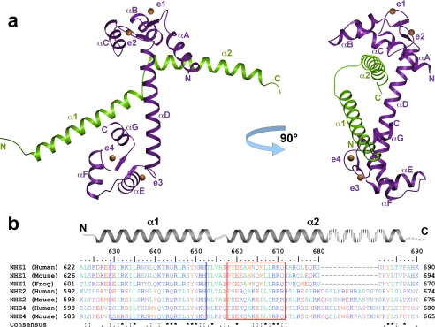
FIGURE 2.
Overall structure of the NHE1CaMBR/Ca2+/CaM complex and sequence alignment of selected mammalian NHE calmodulin binding regions. a, NHE1CaMBR (green) consists of two α-helices (α1 and α2) connected by a short loop. CaM (purple) is present in an elongated form with a central helix (αD) connecting both lobes, which bind to both NHE1CaMBR helices. Each EF-hand of CaM (indicated as e1–e4) binds one Ca2+ ion (orange spheres). b, in the sequence alignment of the crystallized human NHE1 fragment with the corresponding sequences of human NHE2 and NHE4, mouse NHE1, NHE2, and NHE4, as well as frog NHE1, both CaM-interacting regions are marked by a blue and red box.

