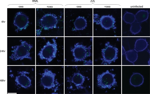
FIGURE 4.
Distribution of “rPrPSc granules” outside and inside cells at various times after infection with RML or 22L, in the absence or presence of swa. PK1 cells were grown for 3 days with or without 2 μg of swa/ml and exposed to a 10−3 dilution of RML or 22L brain homogenate for various times. At 4, 24, and 48 h post-infection, cells were digested with thermolysin to remove PrPC, biotinylated to visualize the cell surface, fixed, and fluorescently stained for rPrPSc (green) and biotin (blue). A thick layer of biotinylated material coated the surface of infected cells, as compared with a faint layer on uninfected cells. External and internal granules of rPrPSc were quantified by confocal imaging (supplemental Table S1). Relatively few rPrPSc granules were internalized, but uptake was not affected by swa. White bar, 10 μm.

