FIGURE 7.
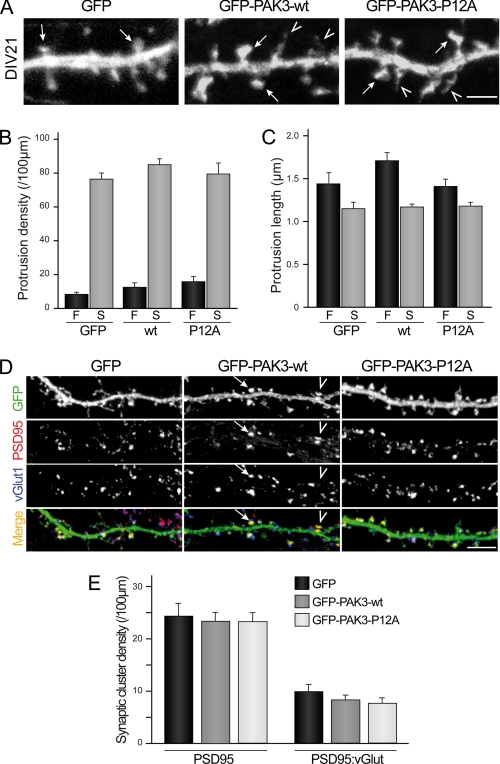
PAK3 acts on spinogenesis and synaptogenesis independently of its interaction with Nck2. A, P12A mutation did not affect spine maturation. Hippocampal neurons were transfected either with GFP as control or GFP-PAK3-WT or GFP-PAK3-P12A constructs at DIV18 and fixed at DIV21. Confocal images showed mature spines (arrows) and filopodia (arrowheads) at DIV21. Scale bar represents 2 μm. B and C, quantitative analysis of protrusion density or length. No significant difference for filopodia (F) or spines (S) between the three expressed constructs was observed. Data are means ± S.E. of measurements obtained from 8 to 11 cells (length of dendrites analyzed: 50–250 μm/cells; 1030–1800 protrusions analyzed). D, P12A mutation did not affect synaptic density. Hippocampal neurons were transfected either with GFP (1st panels) as control, GFP-PAK3-WT (2nd panels) or GFP-PAK3-P12A (3rd panels) constructs at DIV18, fixed at DIV21, and immunolabeled for PSD95 as postsynaptic marker and vGLUT1 as presynaptic marker. Confocal images showed GFP proteins in green, PSD95 in red, and vGLUT1 in blue. PSD95-positive spines were identified from green and red images (arrowhead), and synapses were identified as PSD95-positive spines with adjacent staining of vGLUT1 (arrow). Scale bar, 5 μm. E, quantitative analysis of synaptic cluster density. No significant difference was observed in PSD95-positive spine (left histogram) or synapse (right histogram) density between the three conditions. Data are means ± S.E. of measurements obtained from 15 to 17 neurons (length of dendrites analyzed: 50–250 μm/cell; 680–820 PSD-95 clusters and 230–330 synapses analyzed).

