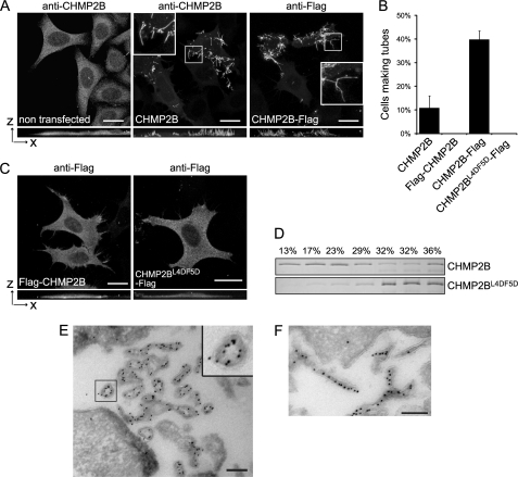
FIGURE 2.
Overexpression of CHMP2B or CHMP2B-FLAG induces formation of long cell surface protrusions in which they concentrate. HeLa cells were transfected with the indicated plasmids and immunostained 36 h later. Except otherwise stated, photographs presented in all figures are maximal intensity projections of confocal image stacks. In A and C, bottom panels represent projections in the x-z plane. Bars, 20 μm. A, CHMP2B immunostaining of cells transfected with the empty vector reveals a homogeneous cytoplasmic localization of the protein. Overexpression of CHMP2B and CHMP2B-FLAG induces the formation of cell surface protrusions in which CHMP2B concentrates. Note the absence of cytoplasmic staining in tube forming cells. B, percentage of transfected cells displaying tubes as revealed with anti-CHMP2B antibodies. For each condition, 200 CHMP2B-expressing cells were counted. Data from three independent experiments are plotted. Standard error bars are shown. C, FLAG-CHMP2B and CHMP2BL4D/F5D-FLAG do not induce surface protrusions. D, flotation experiments demonstrate that mutations in Leu-4–Phe-5 impair the capacity of CHMP2B to associate with membranes in vitro. Purified recombinant full-length CHMP2B and CHMP2BL4D/F5D were mixed with liposomes. Liposomes were floated on a sucrose gradient, fractions were run on SDS-PAGE, and proteins were revealed by Coomassie staining. E and F, FLAG immunogold labeling of cells overexpressing CHMP2B-FLAG. Cross-sections of tubes reveal the presence of the C-terminal FLAG lining the lumen of a hollow, electron-dense structure closely associated with the membrane of tubes; longitudinal sections reveal the presence of CHMP2B along the entire length of the tubes. Scale bars, 200 nm.

