FIGURE 6.
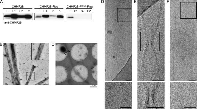
Characterization of tubes pelleted from culture media of CHMP2B-expressing cells. A, Western blotting analysis using anti-CHMP2B of culture media of CHMP2B, CHMP2B-FLAG, and CHMP2BL4D/F5D-FLAG expressing cells. L, cell lysates; P1, 30,000 × g pellet of culture media. P2 and S2, P1 pellets solubilized in 1% Triton X-100 were centrifuged at 20,000 × g: most of the CHMP2B-containing material present in culture media is resistant to detergent extraction. No CHMP2B pelletable material could be recovered from media of CHMP2BL4D/F5D-FLAG-expressing cells. B, P1 pellets of culture medium of CHMP2B-FLAG-expressing cells contain tubes made up of CHMP2B. P1 pellets were fixed, permeabilized, and immunolabeled with anti-CHMP2B antibodies revealed by protein A gold (10 nm). C–F, cryo-EM analysis of P1 pellets prepared from culture medium from CHMP2B-FLAG-expressing cells reveals the structure of CHMP2B tubes. C, low magnification shows tubes with a length reaching at least 8 μm. D, striations perpendicular to the longitudinal axis can be seen across the width of the tube. An asterisk indicates a 50-nm vesicle inside of the tube. E, constriction of a tube with no change in the helix pitch. F, dome closing one end of a tube. The inner leaflet of the membrane is closely associated with the CHMP2B protein lattice. Scale bars, 50 nm.

